Alzheimer Gehirn Ct

Brain Ct Scan Ct Brain Scan From The Top Of Head Affiliate

Mgh Martinos Center On Twitter Brain Art Anatomy Art Medical Art

Creutzfeldt Jakob Disease Cjd Is A Spongiform Encephalopathy

Professor S Brain Soup Experiment Alzheimers Medical

Mesial Temporal Sclerosis Mts Also Commonly Referred To As

Sturge Weber Dimitri Syndrome 1 Brain Images Pathology Medical
This is perhaps most marked in the parietal regions.
Alzheimer gehirn ct. Either like a zumba or taichi or yoga. When they did have an apartment open we moved him from next door to there. Caring104725950 2015 01 20 my father was at an assisted living facility but we kept him going at the day program of the alzheimer s resource center because it was much higher quality. Get easy access to resources community programs and services.
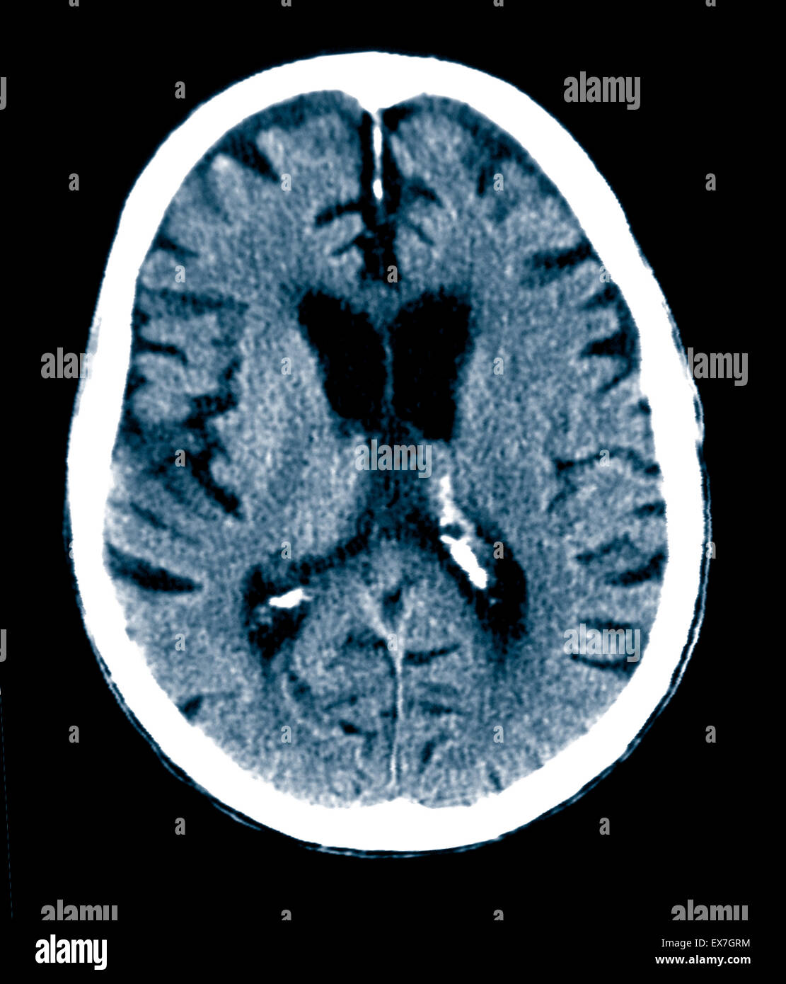
Alzheimer s resource center of connecticut in plantsville ct has an overall rating of 3 out of 5 abd a long term care rating of average. Die großhirnrinde schrumpft und schädigt die bereiche die am denken planen und erinnern beteiligt sind. How a head ct scan can detect alzheimer s disease a head ct scan looks at the structure of your brain. Im hippocampus dem bereich der großhirnrinde der bei der bildung neuer erinnerungen eine wichtige rolle spielt ist die schrumpfung besonders ausgeprägt.
The preferential involvement of the hippocampi and parahippocampal gyri with relative sparing of the rest of the temporal lobe is fairly typical of alzheimer disease or limbic predominant age related tdp 43 encephalopathy late. It is a medium facility with 120 beds and has nonprofit. Unfortunately these two entities can not only mimic each other clinically and. The degree of atrophy in advanced dementia can be striking.
Everyday they have two forms of exercise options. Ct scan shows enlargement of cerebral sulci and loss of gyral volume associated with mild compensatory dilation of the ventricular system. Right now there are 80 000 people living with alzheimer s disease in connecticut and more than 178 000 family members and friends providing care.

Double Cortex Syndrome Radiology Case Radiopaedia Org

Ct And Mri In A Patient With Tuberous Sclerosis There Are

Mount Fuji Sign Is Seen On Cross Sectional Imaging And Implies

Hygiene And Alzheimer S Science News Naked Scientists

Pin On Id

How To Read A Brain Ct Scan Moderate In 2020 Brain Scan Ct Scan
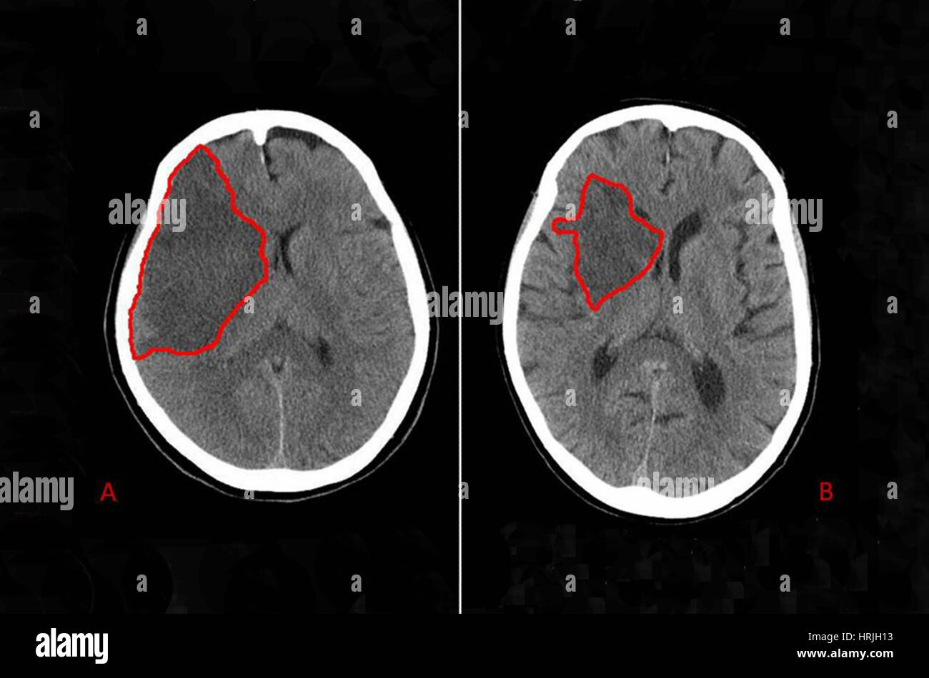
Brain Stroke Ct Scan Stock Photo Alamy

Ct T1 T2 Flair Gre Pd Jpg 512 471 Neurology Radiology

Mastermed Is A Well Known Supplier Of Used Refurbished Mri Scan

Pin Auf Health Beauty Tips
Dementia Brain Scan High Resolution Stock Photography And Images

Normal Brain Anatomy As Demonstrated By Computerized Tomography

Neurofibromatosis Type Ii Radiology Case Radiopaedia Org

Connecticut Hypnotherapy Contact Us Ct Hypnotherapy With

Pineoblastoma Radiology Case Radiopaedia Org Radiology

The Human Brain Laminated Anatomy Chart Human Brain Anatomy

P3320336 Ct Scan Of Brain Spl Jpg 530 436 With Images Brain

